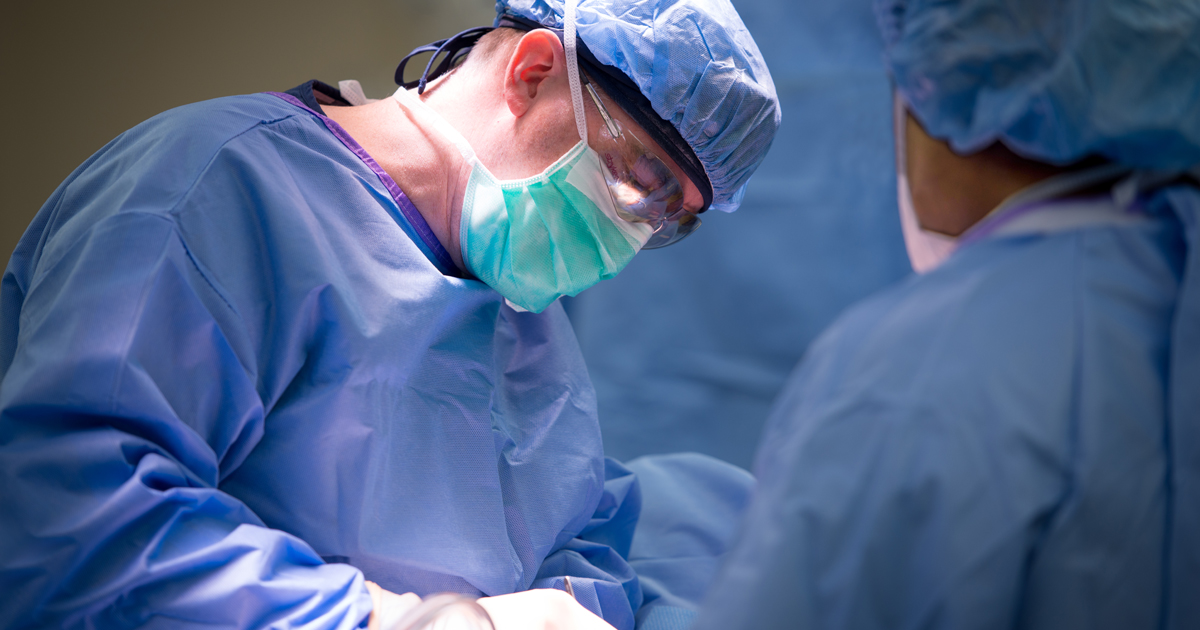
Most patients with a solid tumor undergo surgery to remove the cancer. But many times—20 percent to 60 percent of the time, in fact—surgeons miss something. That's because cancer surgery is challenging, and sometimes imprecise. Surgeons may have difficulty, for example, determining whether and where the cancer has spread. Some also have trouble determining whether small lesions are cancerous or whether all the diseased tissue has been removed. To help improve surgical accuracy, researchers are testing dyes that make cancer cells light up so surgeons can find them more easily, while also allowing them to spare healthy tissue.
"Each year, thousands of patients have to undergo secondary surgeries to remove cancerous tissue that was left behind," says Anita Johnson, MD, FACS, Director of Breast Surgical Oncology at our hospital in Atlanta. "Fluorescence-guided procedures are now being investigated to help reduce the need for a secondary surgery."
Lighting up cells
The idea of using fluorescent dyes to help surgeons differentiate cancerous from benign tissue has been around for some time. About a decade ago, Sunil Singhal, MD, a Penn Medicine physician, became inspired while staring at the fluorescent stars pasted on the ceiling of his baby's room. Thinking "how cool it would be if we could make cells light up," he decided to try the U.S. Food and Drug Administration (FDA)-approved contrast agent, indocyanine green (ICG), to do just that, recognizing that tumor tissue holds onto ICG longer than other tissue. By administering the dye intravenously to patients before surgery, the tumors would glow when lit with a near-infrared camera even after the dye had disappeared from the rest of the body. Dr. Singhal is currently testing the dye, called TumorGlow®, for lung and brain cancer, among other cancer types.
Other dyes are also undergoing testing. One binds to a protein that occurs more commonly in cancer cells than in healthy ones. Another attaches to a molecule that travels to tumor cells. Some dyes are injected into patients a few days before surgery, and others are administered a few hours beforehand. Some of the dyes have gained FDA approval, while others are still being tested in ongoing clinical trials. In addition to being tested on lung and brain cancer, the dyes are also in experimental use in surgeries to remove breast cancer, colorectal cancer, head and neck cancer, and ovarian cancer.
Future of care
Testing has revealed some limitations. For instance, the dyes give false positives in certain cases, with healthy tissue lighting up when it shouldn't. But most surgeons would rather take out a little more healthy tissue during an initial surgery than schedule a second operation to remove cancerous tissue that was missed the first time around, Dr. Johnson says. "As a breast cancer surgeon, it helps to know which breast tissue to remove and which tissue to leave alone," she says. "This form of imagery may reduce the risk of subsequent operations in patients who undergo breast conservation surgery." Right now, up to one-third of women with breast cancer who have a lump removed end up having a second operation.
Many cancer experts believe the dyes will not only be incorporated into most solid-tumor surgeries soon, but also will become an important part of the future of cancer treatment. "Based on the current literature, I think this form of image-guided surgery would be beneficial to patients, as well as to the oncology health care system as a whole," Dr. Johnson says. "More than likely, this will become standard of care within the next decade."


